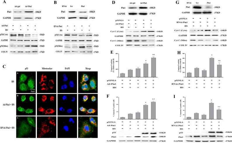
Fig. 5. Pin1 potentiated p53 transcription-independent apoptotic activity after exposure to heat stress (HS).
The p53+/+ mouse aortic endothelial cells (MAECs) were transfected with Ad-Pin1 24 h or Pin1 siRNA 48 h in advance, exposed to 43 °C for 2 h, and then further incubated at 37 °C for 6 h (a–c). a Western blots of Pin1 protein expression in transfected cells and intracellular location of p53 in Pin1-overexpressing p53+/+ MAECs following HS. b Western blots of Pin1 protein expression in transfected cells and intracellular location of p53 was determined by western blots in Pin1 RNAi transfected p53+/+ MAECs following exposure to HS. c Confocal laser scanning microscopy assessment of localization of p53 to mitochondria in Pin1-overexpressing or Pin1 RNAi transfected p53+/+ MAECs exposed to HS. The green fluorescence represents p53, red fluorescence represents Mito Tracker, and orange fluorescence represents translocation of p53 from cytoplasm to mitochondria. H1299 cells were transfected with the p53NLS- construct 48 h and/or Ad-Pin1 24 h in advance. The transfected cells were exposed to 43 °C for 2 h and then further incubated at 37 °C for 6 h (d–f). d Western blots of Pin1 protein expression in transfected cells and the levels of Cyt C in the mitochondrial and cytosolic fractions in Pin1-overexpressing p53+/+ MAECs following HS. e The enzymatic activity of Caspase-9 was measured using fluorogenic substrate Ac-LEHD-AFC. f To quantify induction of apoptosis by HS, Annexin V-FITC/PI staining and flow cytometry were used. Western blots of Pin1 and p53 protein expression in transfected cells. H1299 cells were transfected with the p53NLS- construct and/or Pin1 siRNA 48 h in advance. Cells were exposed to 43 °C for 2 h and then incubated at 37 °C for 6 h (g–i). g Western blots of Pin1 protein expression in transfected cells and the levels of Cyt C in the mitochondrial and cytosolic fractions in Pin1 RNAi transfected p53+/+ MAECs following exposure to HS. h Enzymatic activity of Caspase-9 was measured using fluorogenic substrate Ac-LEHD-AFC. i Induction of apoptosis in response to HS was analyzed by flow cytometry with Annexin V-FITC/PI staining. Western blots of Pin1 and p53 protein expressed in transfected cells. *P < 0.05 compared with the HS group in H1299 p53-/- cells (37 °C); #P < 0.05 compared with the HS group in H1299 p53NLS- cells

