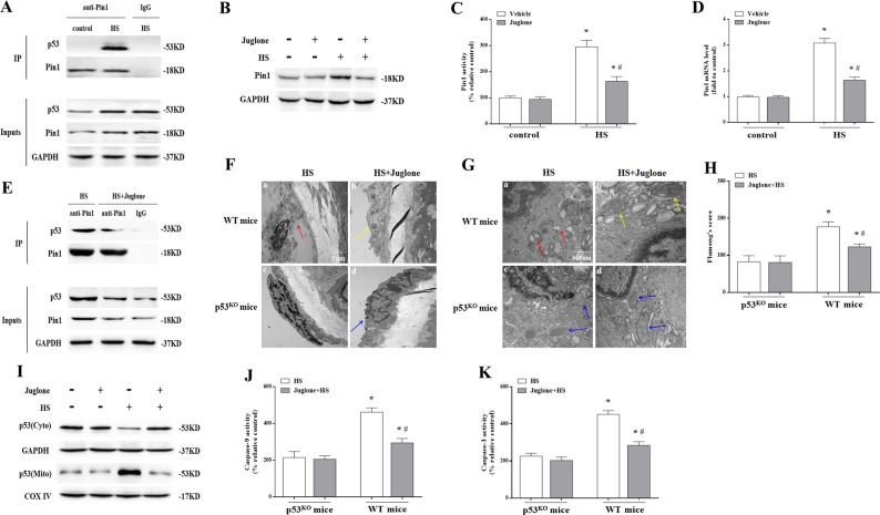
Fig. 6. The effect of Pin1 inhibition on p53 transcription-independent pathway in aortic endothelium induced by heat stress (HS).
Mice were put through the same HS assay as previously described. In the juglone-treated group, each mouse received an intraperitoneal injection of juglone (1 mg/kg body weight/d) for 3 consecutive days prior to exposure to HS. The mice were sacrificed at 6 h after exposure to HS and the aortic endothelium was isolated. a Interaction between endogenous p53 and Pin1 was analyzed by co-immunoprecipitation. IgG served as the negative control. b Pin1 protein levels were assessed by western blot. c Quantification of Pin1 activity in each group. d Quantification of Pin1 mRNA in each group. e Using co-immunoprecipitation to analyze the effect of Juglone on interaction between p53 and Pin1. IgG served as the negative control. f Aortic endothelium morphology was observed using transmission electron microscopy (TEM). f(a) Represents the HS group in wild-type mice and the red arrow indicates damaged endothelial cells; f(b) represents the HS + Juglone group in wild-type mice and the yellow arrow indicates damaged endothelial cells; f(c) represents the HS group in p53KO mice; f(d) represents the HS + Juglone group in p53KO mice and the blue arrow indicates damaged endothelial cells. g Mitochondrial morphology in endothelial cells of aortic endothelium was observed by TEM. g(a) Represents the HS group in wild-type mice and red arrows indicate mitochondria with severe damage, which appear swollen and irregularly shaped with disrupted and poorly defined cristae; g(b) represents the HS + Juglone group in wild-type mice and yellow arrows indicate mitochondria with partial damage, which appear mildly swollen and irregularly shaped and displayed less damage than the wild-type mice. g(c) and g(d) represent the p53KO and p53KO + Juglone group, respectively, and blue arrows indicate mitochondria with partial damage, which appear mildly swollen and irregularly shaped with no significant difference between the two groups, but less damage than in the wild-type mice after exposure to HS. h Mitochondria structural damage was evaluated by Flameng’s score. i Levels of p53 in mitochondrial and cytosolic fractions were assessed by western blot. j Enzymatic activity of Caspase-9 was measured using fluorogenic substrate Ac-LEHD-AFC. k Enzymatic activity of Caspase-3 was measured using fluorogenic substrate Ac-DEVD-AMC. *P < 0.05 compared with the control group in p53KO mice (c, d) or the HS group in p53KO mice (h, j, k); #P < 0.05 compared with the HS group in wild-type mice, n = 6

