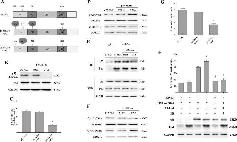
Fig. 7. Phosphorylation of p53 on Ser46 was an important event in Pin1 induction of p53 transcription-independent apoptotic activity in response to heat stress (HS).
H1299 p53-/- cells were transfected with the p53NLS-, p53NLSm S46A, or p53NLSm S46wt constructs 48 h in advance. Cells were exposed to 43 °C for 2 h and then further incubated at 37 °C for 6 h. a Schematic of p53 indicating Pin1 consensus sites (phospho-Ser/Thr-Pro). TA represents transactivation domain, PRD represents proline enrichment domain, DBD represents DNA binding domain, and NLS represents nuclear localization signal. The p53NLS mutants had Ser/Thr-to-Ala substitutions at the Pin1 consensus sites at residue 46 (p53NLSm S46A) and two other major Pin1-binding sites at residues 33 and 81 ((p53NLSm S46wt). b Expression of P-Ser 46 p53 and p53 were determined by western blot. c Quantification of the P-Ser 46 p53 and p53 ratio. d Levels of p53 in the mitochondrial and cytosolic fractions were assessed by western blot. e Cell lysates normalized for p53 protein levels were subjected to co-immunoprecipitation to analyze interaction between endogenous p53 and Pin1. IgG served as the negative control. f The levels of Cyt C in the mitochondrial and cytosolic fraction were assessed by western blot. g Quantification of apoptosis in response to HS was performed with flow cytometry and Annexin V-FITC/PI staining. h H1299 p53-/- cells were transfected with the p53 NLS- S46A construct 48 h and/or Ad-Pin1 24 h in advance. Cells were exposed to 43 °C for 2 h and then further incubated at 37 °C for 6 h. The levels of p53 and Pin1 proteins were measured by western blot. Apoptosis in response to HS was quantified by flow cytometry using Annexin V-FITC/PI staining. *P < 0.05 compared with the HS group in H1299 p53NLS- cells; #P < 0.05 compared with the HS group in Ad-Pin1 + H1299 p53NLS cells

