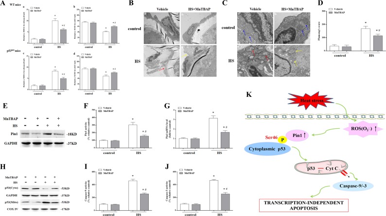
Fig. 9. The effect of inhibition O2−. on Pin1/p53 transcription-independent apoptosis pathways in aortic endothelium following exposure to heat stress (HS).
Mice were put through the same HS assay as previously described. In the MnTBAP-treated group, each mouse received an intraperitoneal injection of MnTBAP (10 μg/g body weight) 1 h prior to HS. The mice were sacrificed at 6 h after exposure to HS and the aortic endothelium was isolated. a Quantification of malondialdehyde (MDA) and superoxide dismutase (SOD levels in wild-type and p53KO mice. b Aortic endothelium morphology was observed by transmission electron microscopy (TEM) in wild-type mice. b(a) and b(b) represent the control and the control + MnTBAP group, respectively; b(c) represents the HS group and the red arrow indicates damaged endothelial cells; b(d) represents the HS + MnTBAP group and the yellow arrow indicates damaged endothelial cells. c Mitochondrial morphology in aortic endothelial cells was observed by TEM in wild-type mice. c(a) and c(b) represent the control and control + MnTBAP group, respectively, and blue arrows indicate mitochondrial morphology, which appeared normal with preserved membranes and cristae; c(c) represents the HS group and red arrows indicate mitochondria with severe damage, which appeared swollen and irregularly shaped with disrupted and poorly defined cristae; c(d) represents the HS + MnTBAP group and yellow arrows indicate mitochondria with partially damage, which appeared mildly swollen and irregularly shaped and had less damage than the wild-type mice exposed to HS. d Mitochondria structural damage was evaluated by Flameng’s score. e The levels of Pin1 protein were assessed by western blot. f Quantification of Pin1 activity in each group. g Quantification of Pin1 mRNA in each group. h The levels of p53 in the mitochondrial and cytosolic fraction were assessed by western blot. i Enzymatic activity of Caspase-9 was measured using fluorogenic substrate Ac-LEHD-AFC. j Enzymatic activity of Caspase-3 was measured using fluorogenic substrate Ac-DEVD-AMC. k ROS (O2−.) mediated HS-induced apoptosis in vascular endothelial cells through a Pin1/p53 transcription-independent pathway. *P < 0.05 compared with the control group; #P < 0.05 compared with the HS group, n = 6

