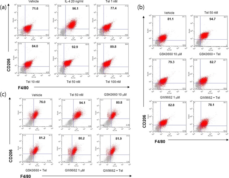
Figure 2.
FACS analysis of telmisartan-induced M2 polarization. RAW264.7 macrophages (1 × 10⁴ cells/well) were treated with various concentrations (1, 10, 50 and 100 nM) of telmisartan for 24 h, and then flow cytometry was conducted using CD206 and F4/80 antibodies (a). RAW264.7 macrophages (b) or primary BMDM (c) were pretreated with PPARγ antagonist (GW9662, 1 μM) or PPARδ antagonist (GSK0660, 10 μM) for 1 h, and then treated with telmisartan (50 nM). Changes in the M2 population were determined by flow cytometry. IL-4 (20 ng/ml) was chosen as an M2 inducer. Representative results are shown.

