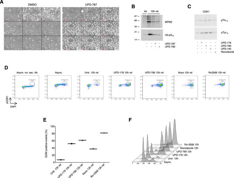
Figure 4.
CDC25 inhibitors impair the execution of mitosis. (A) Phase contrast stills of HeLa cells synchronized by 2x thymidine block-release, treated with vehicle alone or UPD-787 (10 μM) 5 h upon release and visualized from 7.5 h to 21.5 h upon release (4 frames/h). (B) Western blot analysis of MPM2 epitopes and Histone H3 phosphorylation (pSer10) in HeLa cells treated as in (A) and examined at 10 h upon release from the 2x thymidine block. (C) Western blot analysis of CDK1 (pThr14 and Tyr15, respectively) from HeLa cells treated as in (A) and examined at 10 h upon release from the 2x thymidine block. (D) Flow cytometric analysis of CDK1-pTyr15 in cells treated as in (A) and examined 12 h upon release from the 2x thymidine block. Nocodazole (0.4 μg/ml) and Ro-3306 (9 μM) were used as controls for the level of CDK1 phosphorylation. Staining with secondary antibody only (left panel) was used to subtract self-fluorescence. (E) Quantification of positive events gated as shown in (D). (F) DNA content (DAPI staining) of the cells shown in D.

