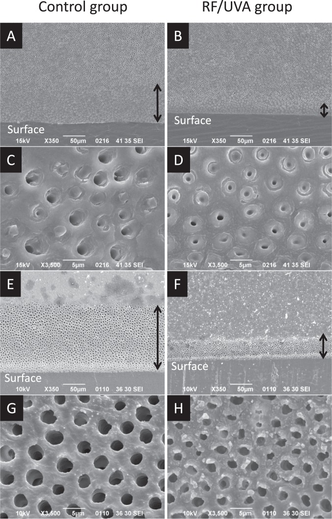
Figure 3.
Scanning electron microscope images of demineralized dentin surfaces. Samples were immersed in a demineralizing solution of pH 5.0 (A–D) and 10% ethylenediaminetetraacetic acid (EDTA) (E–G) for 3 days each. (A,C,E,G) control group; (B,D,F,H) riboflavin/ultraviolet light (RF/UVA) treated group. The depth of the demineralized zone (black arrow) was clearly larger in the control (A,E) compared to that in the RF/UVA group (B,F). Dentinal tubules in the demineralized zone were enlarged in the control group (C,G), while the demineralization was less aggressive in the RF/UVA group (D,H).

