Figure 5.
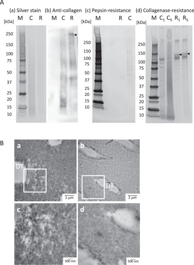
Enzymatic resistance of dentin collagen after riboflavin/ultraviolet light (RF/UVA) treatment. (A) SDS-PAGE and western blotting of dentin collagen after RF/UVA treatment. M: marker, C: control group, R: RF/UVA treatment group, C1, C5: control group, treated by collagenase for 1 or 5 days, R1, R5: the RF/UVA group, treated by collagenase for 1 or 5 days. The silver staining shows there was no difference in total protein between the control and the RF/UVA groups (a). The anti-collagen antibody staining shows that type-I collagen with high molecular weights were present in the RF/UVA group (black arrow), whereas collagen with lower molecular weights was predominant in control group (b). After treatment with pepsin, the resulting profile shows that dissolved collagen was observed in the control group, while no clear profile appeared in the RF/UVA group (c). After treatment by collagenase for 1 and 5 days, the profiles show that dissolved collagen were detected in the control group (d). After 5-day treatment by collagenase, highly dissolved collagen appeared as a contiguous belt in the control group, while the collagen profiles after RF/UVA treatment showed clear bands in the area of 100–120 kDa. Full-length gels and blots are presented in Supplementary Figure S1. (B) TEM images of dentin specimens degraded by collagenase for 5 days. a and c: control group; b and d: RF/UVA group. Areas indicated with white squares in a and c are shown magnified in c and d. DT: dentinal tubules. The collagen around dentinal tubules was severely degraded in the control group after degradation (a,c), whereas in the RF/UVA group, the collagen retained its original form (b,d).

