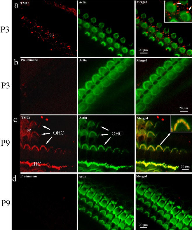
Figure 2.
Immunofluorescent confocal imaging of organ of Corti from the apical cochlear region at P3 and P9. (a) TMC1 is not detected on stereocilia at P3, although the kinocilium appears labelled (arrow in merged image, inset), and speckled labelling is noted on supporting cells. Phalloidin confirms the presence of hair bundles at the same focal plane. (b) Pre-immune serum did not generate any labelling at P3. (c) TMC1 labelling was detected in IHC and OHC bundles at P9; colocalisation with stereocilia is observed as a yellow colour in the merged image. Weak fluorescence was detected on the hair-cell apical surface. The inset shows an enlargement that reveals red and green signal in the indicated (arrow) bundle can also be distinguished. (d) Pre-immune serum did not produce labelling. In all red channel images, the green channel was subtracted using the confocal software to remove bleed through from the phalloidin staining.

