Fig. 3.

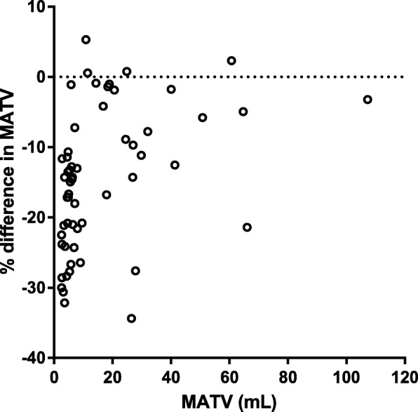
Relative difference (%) in lesion MATV (mL) between uncorrected and PVC images (LR + HYPR) as function of MATV on uncorrected images. Y-axis was scaled to − 40%; for one lesion of 5.8 mL MATV was 69% smaller on PVC image

Relative difference (%) in lesion MATV (mL) between uncorrected and PVC images (LR + HYPR) as function of MATV on uncorrected images. Y-axis was scaled to − 40%; for one lesion of 5.8 mL MATV was 69% smaller on PVC image