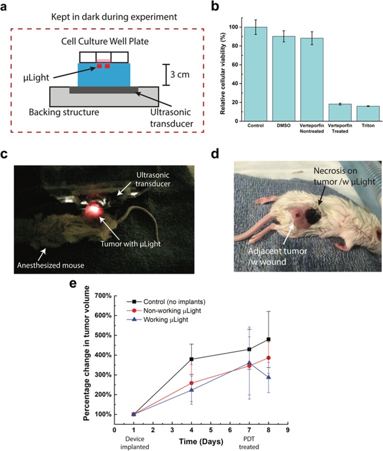
Figure 3.
(a) In vitro experimental setup, (b) cytotoxicity assay of experimental groups: control (no treatment), cell culture media with ultrasonic treatment; DMSO only; verteporfin added but non-treated; expectation (verteporfin introduced cell culture media with active μLights by ultrasonic treatment); and Triton X100 treated (added to kill cells), (c) in-vivo experimental setup: μLight was implanted in mouse and excited/powered via ultrasound, (d) optical image of the mouse (24 hours after PDT treatment), (e) Tumor volume change with respect to the size when device was implanted. PDT treatment with μLights was conducted 7 days after the surgery allowing for wound healing.

