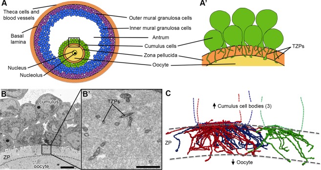
Figure 1.
Cumulus cells send numerous projections through the zona pellucida. (A) Schematic of a mouse antral follicle. Black square represents area magnified in (A’). (A’) Schematic of several cumulus cells that send transzonal projections (TZPs) through the zona pellucida to connect to the oocyte. Cumulus cell bodies can be adjacent to or displaced from the zona pellucida. (B) SEM image of a cross-section through cumulus cells, zona pellucida (ZP, outlined by dashed lines), and oocyte of an antral follicle. Video 1 shows 202 serial sections of this area and highlights a cumulus cell with all of its projections. Scale bar, 5 μm. (B’) High-magnification of a region in the zona pellucida showing several TZPs in cross-section imaged by SEM. Scale bar, 1 μm. (C) Reconstruction of every TZP sent by three neighboring cumulus cells, each shown in a different color. TZPs were segmented from 405 serial electron micrographs, encompassing a volume of 22.5 × 6.7 × 18.2 μm (x, y, z). ZP, zona pellucida.

