Figure 3.
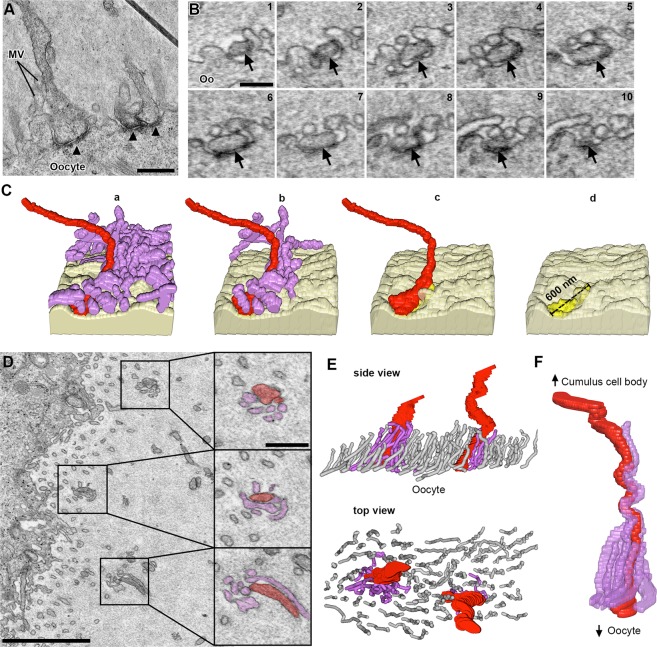
Contacts between TZPs and oocyte components. (A) TEM image of TZPs that make adherens junctions with the oocyte surface. Adherens junctions are identified by an electron-dense region at the site of TZP-oocyte contact (black triangles). Mv, oocyte microvilli. Scale bar 500 nm. (B) Serial section SEM images of a TZP (black arrow) that makes an adherens junction with the oocyte surface. Oo, oocyte. Scale bar, 300 nm. (C) Reconstruction of an adherens junction made by the TZP shown in B. Reconstruction is 2.0 × 1.1 × 1.7 μm (x, y, z), spanning through 38 serial sections (each, 45 nm-thick). Light yellow: oocyte surface. Purple: oocyte microvilli. Red: TZP. Bright yellow: adherens junction. (a) TZP and all oocyte microvilli in the volume. (b) Unattached microvilli have been removed from the reconstruction to show only those that make a contact with the TZP. (c) All microvilli have been removed from the reconstruction. (d) TZP has been removed from the reconstruction to show the adherens junction on the oocyte surface. (D) SEM image of an area of the zona pellucida in which TZPs and microvilli appear clumped (squares). High-magnification subpanels show the TZP in red and oocyte microvilli in purple (confirmed by serial sections). Scale bars, 2 μm on low magnification, and 500 nm on high magnification subpanels. Video 5 shows this in serial sections (reconstructed in F). (E) Side and top views of a reconstruction of an area in the zona pellucida that is 3.4 × 1.8 × 2.8 μm (x, y, z), spanning through 63 serial sections (each, 45 nm-thick), showing two areas where TZPs (red) are seen clumped with microvilli from the oocyte (purple). Non-interacting microvilli are shown in gray. (F) Reconstruction of a single TZP (red), and oocyte microvilli that tightly contact it (purple). The reconstruction is 2.9 × 1.2 × 2.7 μm (x, y, z), spanning through 61 serial sections (each, 45 nm-thick). This reconstruction was segmented from the data seen in Video 5.

