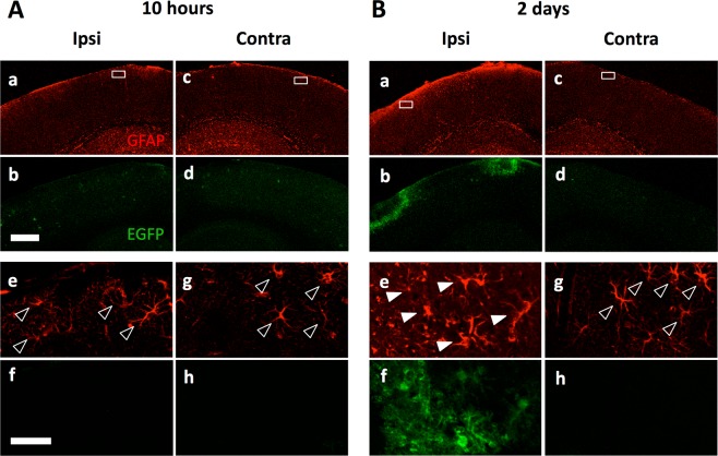
Figure 1.
Astrocytes were activated by the cranial window operation on the GFAP-EGFP mice brain. Immunohistochemistry for EGFP (green) and GFAP (red) expression in cerebral cortex 10 hours (A) and two days (B) after craniotomy in one side (ipsilateral; Ipsi). The contralateral sides (Contra) are shown as a control (Ac,d,g and h). (Ae–h and Be–h) Higher magnifications of the boxes in top panels are shown. GFAP-driven EGFP was only detectable in the operated side of cortex with delay of more than one day. Filled arrowheads and open arrowheads indicate EGFP-positive/GFAP-positive astrocytes and EGFP-negative/GFAP-positive astrocytes, respectively. Scale bar, 500 µm (Ab) and 50 µm (Af).

