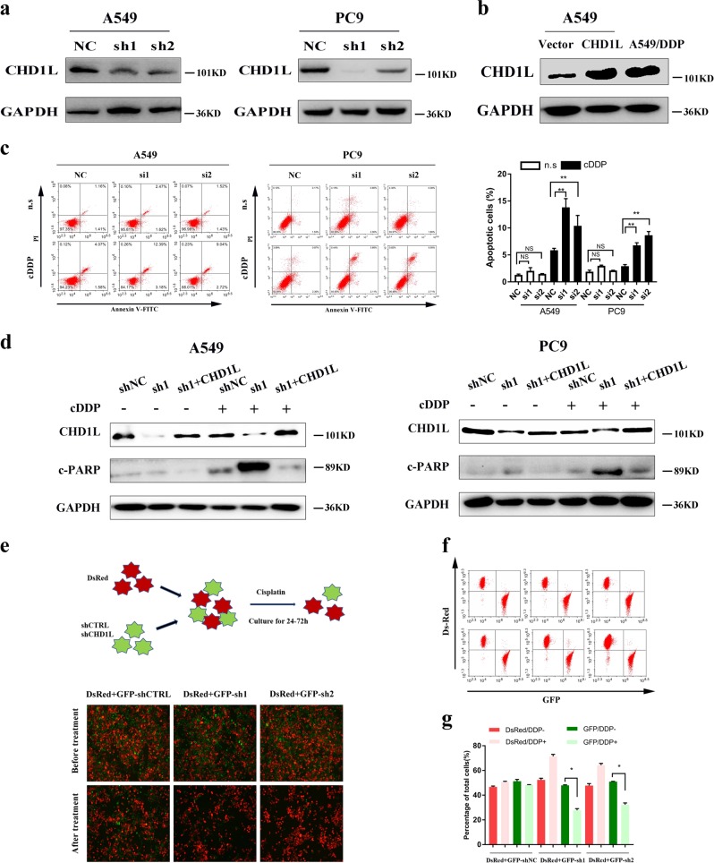
Fig. 2.
CHD1L suppresses cisplatin-induced apoptosis in NSCLC cells. a Western blotting reveals that CHD1L was efficiently knocked down by the treatment of CHD1L-shRNA-1 or CHD1L-shRNA-2 in A549 and PC9 cells. b Protein expression of CHD1L in vector, CHD1L-overexpressed (CHD1L) and A549/DDP cells. c Annexin-V-FITC/PI dual staining assay (left, representative plots for flow cytometry; right, bar charts indicating the average percentages of apoptotic cells). d WB assays (c-PARP, cleaved PARP; GAPDH, a loading control). e A549 cells stably transfected with shRNAs specifically targeting CHD1L or control shRNAs were labeled with GFP; meanwhile, the control parental cells were labeled with DsRed. GFP positive and DsRed positive cells were mixed together and subjected to cisplatin treatment (10μM) for 24-72 hours. f Fluorescent images of A549-shCTR-GFP and A549-shCHD1L-GFP cells mixed with A549-DsRed cells before and after cisplatin treatment. g Statistical analysis of flow cytometry data. NC, negative control siRNA; si1, si2, CHD1L siRNAs; Vec, empty vector transfected; CHD1L, CHD1L overexpression; n.s, normal saline. *P<0.05; **P<0.01; NS, no significance

