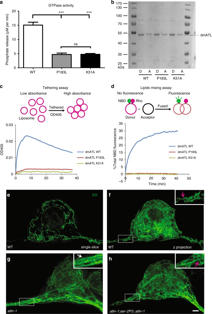
Fig. 2.
ATLN-1 regulates somatic ER network formation. a Quantification of GTPase activity of WT and mutant dmATLs. The activities were measured by phosphate release at saturating GTP concentrations of 0.5 mM. At least three repetitions were performed for each group. Values are mean and error bars are SEM. ns, not significant; ***p < 0.001 (Tukey’s multiple comparison test). b Reconstitution of Drosophila WT, P183L, and K51A dmATL proteins into liposomes. Donor (D) and acceptor (A) vesicles with recombinant proteins were analyzed by SDS/PAGE and Coomassie blue staining. (c) Schematic illustration of liposome membrane tethering assay (upper panel). The clustering of Drosophila WT and mutant dmATL containing proteoliposomes (protein:lipid ratio 1:2,000) was measured by the light absorbance at a wavelength of 405 nm (lower panel). d Schematic illustration of lipid mixing assay (upper panel). Drosophila WT and mutation dmATL were reconstituted at equal concentrations into donor and acceptor vesicles. GTP-dependent fusion of donor and acceptor vesicles was monitored by the dequenching of an NBD-labeled lipid in the donor vesicles (lower panel). Fusion was initiated by addition of GTP. e Representative 3D-SIM image of PVD somatic ER in a WT worm (single focal plane). f–h Representative 3D-SIM maximum-intensity-projection images of PVD somatic ER in WT (f), atln-1 (g) and atln-1; ser-2P3::atln-1 worms at adult stage. Insets show 2X magnified views. The magenta arrows indicate three-way junction and white arrows indicate parallel ER tubules. Scale bar, 1 μm

