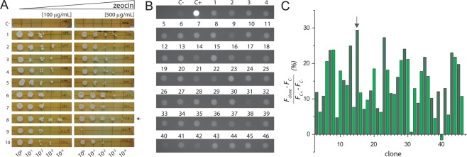
Figure 2.
Clone selection. (A) Drop test in YPD plates with 100 µg/mL and 500 µg/mL of zeocin after 2–3 days of growth at 30 °C. On the left, plates with 100 µg/mL zeocin and, on the right, plates with 500 µg/mL zeocin. Each plate presented a non-transformed SMD1168H serial dilution as negative control (C−). Each number represents a tested clone and (C−) a non-transformed SMD1168H colony. A dilution factor of 10x was done for each lane, starting from left to right. (B) MM plates after 48 hours at 30 °C. Each dot represents a different tested clone from pP-hSGLT1-eGFP transformation except the controls (C−) and (C+). Negative control (C−) is a non-transformed SMD1168H P. pastoris colony. Positive control (C+) is a transformed colony of pP-eGFP empty vector. (C) Densitometry values of tested colonies are expressed in relative fluorescence units (RFU).

