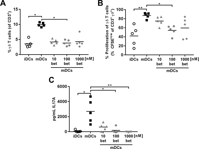
Figure 3.
Dendritic cells (DCs) differentiated with betamethasone impair autologous γδ+ T lymphocyte proliferation. DCs were loaded with insulin (20 µg/ml) and cultured with splenocytes in a ratio 1:10 for 4 days. White circles represent immature DCs (iDCs), black squares represent mature DCs (mDCs) generated in basal conditions and stimulated with lipopolysaccharide (LPS), grey symbols represent mDCs with 10 nM (triangles), 100 nM (dots) and 1000 nM (rhombus) betamethasone (bet) during differentiation and stimulated with LPS. (A) Percentage of γδ+ T cells within CD3+ population after co-culture with DCs. (B) Percentage of autologous proliferation of γδ+ T cells (% CFSElow) after co-culture with DCs. (C) IL-17A production in T cell proliferation experiments measured in supernatants after 4 days of co-culture of DCs and splenocytes. Lines show the mean of 5 independent experiments (*p ≤ 0.05, **p < 0.01, Dunn’s test, Kruskal-Wallis test).

