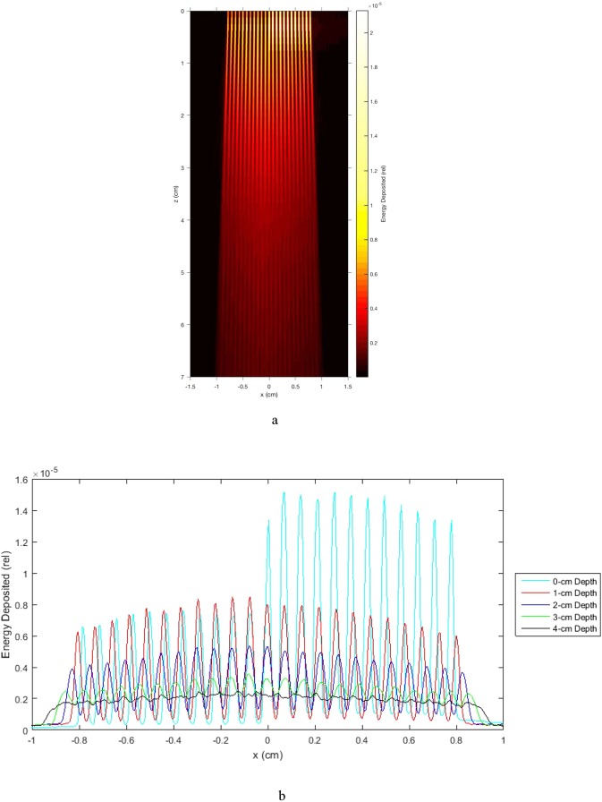
Figure 10.
Effect of cortical bone on the OXM merge depth in water. (a) Shows Monte Carlo simulations of a heatmap of energy deposition by 320 kVp (3.8-mm Cu-HVL) xray minibeams, 300-micron, spaced 700 microns on center, 5-mm-thick tungsten multislit collimator with parallel blades, with a simulated 5-mm slab of cortical bone (density = 1.85) at x > 0 and 0 < z < 0.5 cm. Increased energy deposition is seen in the bone region and a slightly decreased intensity of minibeams is seen in water at x > 0 and z > 0.5 cm, due to attenuation in the bone. Nevertheless, this attenuation led to minimal effect on the merge depth of the minibeams, which occurs at approximately 4-cm depth in water. (b) Shows the horizontal profiles of (a) at depth intervals of 0, 1, 2, 3, and 4-cm for the entire horizontal width of the array. As indicated in the description of (a), the profiles vividly demonstrate that a 5-mm bone has a large effect on the attenuation of x-rays, while it has almost no effect on the merging depth of the minibeam array. The latter is clear from the fact that the profile at the 4-cm depth is completely flat from left to right.

