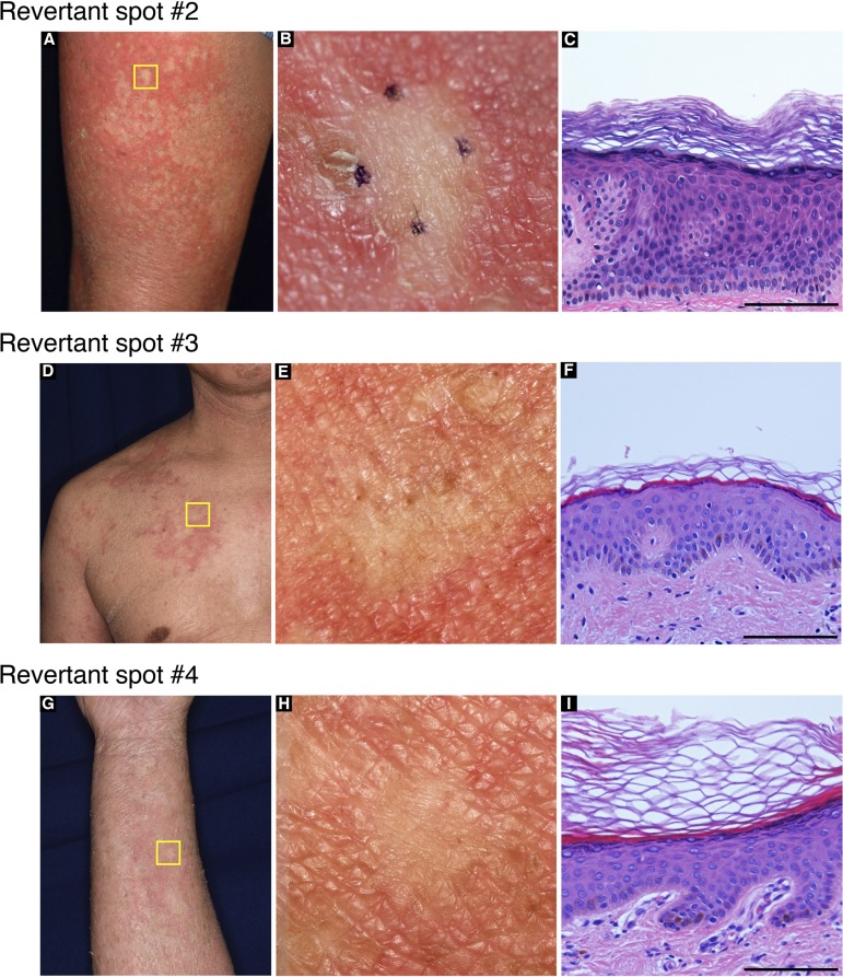
Figure S2. Clinical and histological features of revertant spots in the proband in family 1.
Skin biopsy was taken from multiple smooth-surfaced whitish spots (A, D, G). Higher-magnification images of the squares in (A), (D), and (G) are presented in (B), (E), and (H), respectively. These spots were histologically normalized (C, F, I). Scale bar, 100 μm.

