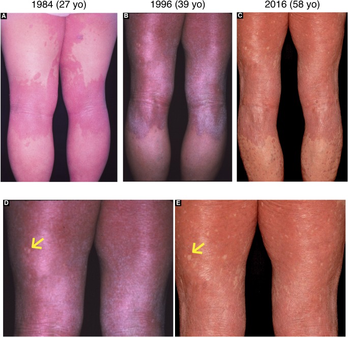
Figure S5. Long-term observation of revertant spots in the proband in family 2.
(A–C) Clinical images of the posterior lower extremities of the proband in family 2, which were taken when she was 27 (A), 39 (B), and 58 (C). (D, E) Higher magnification of her posterior thighs in (B) and (C) demonstrated clinically reverted patches that had increased in both size and number with age. Arrows indicate the largest normal-appearing patch that had been maintained for at least 19 yr on her left posterior thigh.

