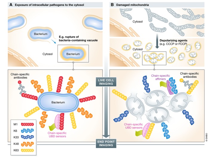
Figure 3. Imaging ubiquitination in selective autophagy.

Schematic overview of live‐cell and end‐point Ub imaging applications in antibacterial autophagy (xenophagy) and autophagic breakdown of damaged mitochondria (mitophagy). (A) Intracellular pathogens, like Salmonella, reside mostly in Salmonella‐containing vacuoles. In some cases, vacuolar membrane rupture and pathogens are exposed to the host cytosol, inducing prominent multi‐type Ub deposition at the bacterial surface. These Ub structures serve as non‐self “eat me” signals that trigger bacterial autophagy. (B) Damaged mitochondria (grey circles), for example induced by depolarizing agents like CCCP/FCCP, trigger PINK1/PARKIN‐dependent ubiquitination at the surface of these organelles, thereby recruiting the autophagy machinery. In both examples, Ub chain‐specific antibodies, UBD‐based chain‐specific sensors and chain‐specific affimers were applied to understand the mechanisms and functional relevance of ubiquitination. Substantial overlap in similarities of Ub patterns exists in these two mechanistically distinct forms of ubiquitin‐accumulated structures and the contribution of Ub for selective autophagy.
