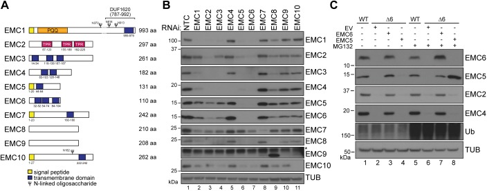
Fig. 1.
EMC5 and EMC6 are essential for EMC maturation. (A) Schematic representation of the primary structure of all EMC subunits (EMC1–EMC10). Domains, boundary residue numbers and predicted glycosylation sites are indicated. Pyrrolo-quinoline quinone (PQQ) and tetratricopeptide repeats (TPR) are shown. (B) siRNA-mediated depletion of EMC1–EMC10 and non-targeting control (NTC) for 72 h in U2OS Flp-In™ T-Rex™ cells. Whole-cell lysates (WCL) of individually depleted cells were separated by SDS-PAGE and resulting western blots probed for each subunit and tubulin (TUB) as indicated. The asterisk (*) denotes a nonspecific band. (C) U2OS Flp-In™ T-Rex™ cells modified by CRISPR/Cas9 to knockout EMC6 (Δ6) were reconstituted by inducing expression of an empty vector control (EV), EMC5 or EMC6 (DOX 1 ng/ml, 72 h). MG132 was added to cells where indicated (5 µg/ml, 8 h). Samples were prepared as in B. TUB, tubulin; Ub, ubiquitin.

