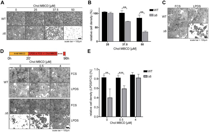
Fig. 2.
EMC-deficient cells are sensitised to cholesterol surplus and starvation. (A) WT and ΔEMC6 (Δ6) cells were exposed to Chol:MBCD (25, 37.5 and 50 µM, 20 h) and visualised by staining with Crystal Violet. (B) Quantification of cell densities for experiments as in A normalised to untreated cells (0 µM Chol:MBCD). Means±s.d. are shown (n=3). **P≤0.01 (Student's t-test). (C) WT and Δ6 cells depleted of cholesterol by MBCD (4 mM, 20 min) were switched to FCS- (5%) or LPDS- (5%) containing growth media (96 h) and visualised by staining with Crystal Violet. (D) Cells were treated as in C with or without Chol:MBCD (0.5 or 4 µM). (E) Quantification of cell density from experiments shown in D. Means±s.d. are shown (n=4). **P≤0.01, ***P≤0.001 (Student's t-test).

