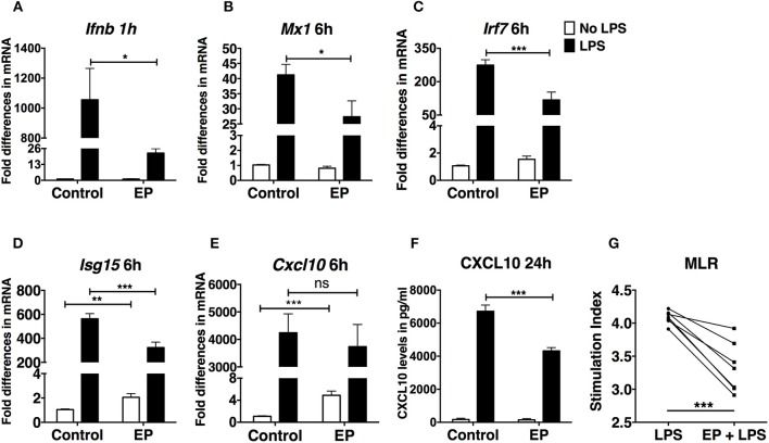
Figure 3.
EP decreases the IFN-I response in LPS-stimulated DCs. DCs were pre-treated with 10mM EP for 1 h before LPS 100 ng/ml. Cells were harvested after (A) 1 h for Ifnb level of expression and (B–E) 6 h for the IFN-I-stimulated gene expression analysis by real-time RT-PCR. Values were expressed as fold difference in mRNA from the control (untreated cells). (F) CXCL10 ELISA from 24 h culture supernatants. Results in (A) are the average of biological replicates from 1 representative of 5 independent experiments (n = 5). Results in (B–E) are from 10 independent experiments (n = 10) and in (F) from 5 independent experiments (n = 5). Results are expressed as mean ± SEM. (G) EP reduces the ability of DCs to stimulate an allogeneic T cell response in the MLR assay. We treated DCs with LPS ± EP pre-treatment for 24 h, then we washed and used them as APCs to induce lymphocyte proliferation in an in vitro MLR assay. We used a DC:splenocyte ratio of 1:30. Lymphocyte proliferation was measured by 3H thymidine incorporation. Stimulation index was calculated by dividing the counts per minute (cpm) for each condition by the cpm of lymphocytes exposed to untreated DCs. Results are from 3 to 4 biological replicates of 2 independent experiments (n = 2, 7 data numbers). Data was analyzed using the two-tailed unpaired Student's t-test. *P < 0.05, **P < 0.01, ***P < 0.001, and ****P < 0.0001.

