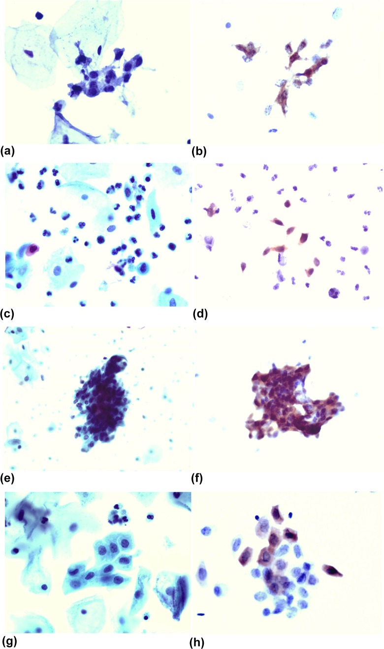
Figure 1.
(a) A slide prepared by the LBP method, showing a cluster of small cell type HSIL cells that are easily overlooked (Papanicolaou stain, ×400). (b) A slide from the same patient immunostained with p16, showing that the HSIL cells are easy to interpret because of the obviously positive nucleus. (c) A slide prepared by the LBP method, showing some scattered HSIL cells that are easily overlooked (Papanicolaou stain, ×400). (d) A slide from the same patient immunostained with the p16, showing that the HSIL cells are easy to interpret because the nucleus is obviously positive. (e) A slide prepared by the LBP method, showing a cluster of HCGs-type HSIL cells that are easily misinterpreted (Papanicolaou stain, ×400). (f) A slide from the same patient immunostained with the p16, showing that the HSIL cells are easy to interpret because the nucleus is obviously positive. (g) A slide prepared by the LBP method, showing a cluster of metaplastic-type HSIL cells that are easily misinterpreted (Papanicolaou stain, ×400). (h) A slide from the same patient immunostained with the p16, showing that the HSIL cells are easy to interpret because the nucleus is obviously positive.

