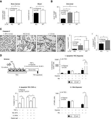
Figure 6.
Intrarenal Mø proliferation and Mø-mediated TEC apoptosis are suppressed in IL-34 KO MRL-Faslpr mice. (A) Proliferation of myeloid progenitors in BM (left panel) and myeloid cells in peripheral blood (right panel) of IL-34 KO and WT MRL-Faslpr mice at 5 months of age analyzed by FACS. (B) Intrarenal Mø proliferation quantified by dual staining for Ki-67 and F4/80 in IL-34 KO, WT, and +/-MRL-Faslpr mice (5 months of age). (C) Intrarenal apoptosis identified using caspase-3 in IL-34 KO, WT, and +/- MRL-Faslpr mice at 5 months of age. Arrows, caspase-3+ cells. 1. Representative photos of tubules and interstitial infiltrates. Graph, analysis of ten HPFs/sample. 2. TEC apoptosis. Quantitation of caspase-3+ e-cadherin+ cell number analyzed by FACS. Dotted line in (B and C) indicates mean at 1.5 months of age. (D) Scheme for in vitro analysis of TECs cocultured with Mø under hypoxic conditions (1% O2 for 48 hours) or with TNF-α (25 ng/ml for 18 hours). 1. Hypoxia: Apoptotic TECs (Annexin V+ CD45−) analyzed by FACS. 2. TNF-α (25 ng/ml for 18 hours): Apoptotic TECs analyzed by toxilight assay. 3. Hypoxia: Mø (F4/80+) number after coculture with TECs analyzed by FACS. Data are mean±SEM. Mann–Whitney U test was used for statistical analysis.

