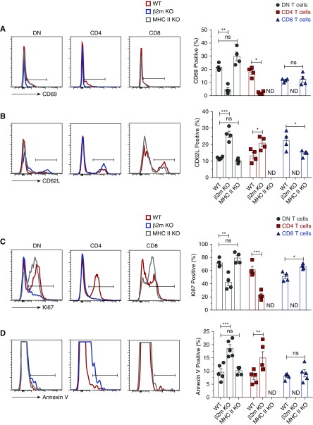
Figure 2.
Lack of β2m, but not MHC II, impairs activation, proliferation, and increased apoptosis of kidney DN T cells in the steady state. Lymphocytes were isolated from kidneys of age-matched WT, β2m KO, or MHC II KO mice, stained, and acquired by LSRII. Gated CD4, CD8, and DN T cells were analyzed for the expression of CD69, CD62L, Ki-67, and Annexin V. Representative plots show percentage of (A) CD69, (B) CD62L, (C) Ki-67, and (D) Annexin V by each T cell type in the kidneys of individual genotype. Graphs show cumulative data from at least four or five independent experiments (n=4–5 mice). Data are expressed as mean±SEM. *P<0.05; **P<0.01; ***P<0.001.

