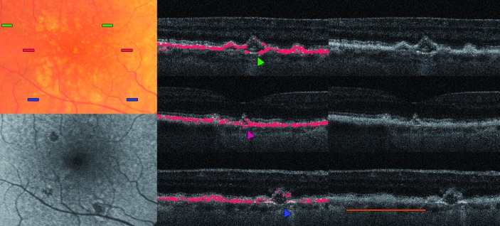
Figure 4.
Eye of a 70-year-old woman showing concave drusen (arrowheads). The colour bars on the fundus photography (top left image) indicate the position of the respective B-scans. In the fundus autofluorescence (below), the retinal pigment epithelium (RPE) hypoautofluorescence can be seen. The orange bar in the bottom right image shows a cluster of indifferent drusenoid lesions with irregular RPE coverage, reminding the saw-toothed pattern.

