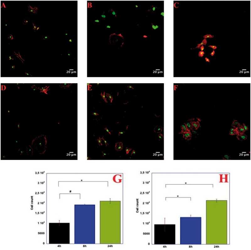
Figure 5.
Confocal Microscopy images of HUVECs on gelatin coated dishes (A, B and C) and the respective histogram with adhesion of cells as a function of seeding time (G). HUVECs on Silk-ELRs co-recombinamer scaffolds (D, E and F) and the related histogram with adhesion of cells as a function of seeding time (H). Scale bar 20 µm. P–values < 0.001

