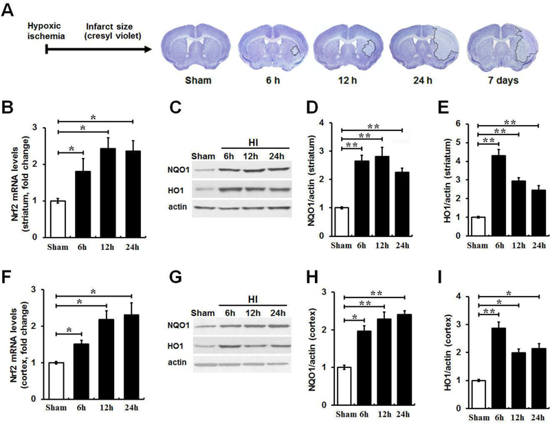
Figure 1. Induction of Nrf2 signature in ischemic brain regions at various times following HI.

(A) The time-dependent ischemic brain damage over 7 days after HI was assessed by cresyl violet staining of coronal brain sections. HI resulted in a rapid increase in infarct size in striatum and cortex within 24 h followed with a gradual and spontaneous recovery on day 7. The initial ischemic damage occurred in striatum and then spread to nearby brain regions. (B and F) qRT-PCR was used to determine the Nrf2 mRNA level in the ischemic striatum and cortex at 6, 12 and 24 h following HI. Nrf2 mRNA levels had a 1.5–2.4 fold increase within 24 h after HI in both brain regions compared to sham controls, whereas there is no significant difference between indicated time points. (C, D, E, G, H and I) Western blot results were quantified for the protein levels of Nrf2 target markers NQO1 and HO1 in ipsilateral ischemic striatum and cortex at 6, 12 and 24 h following HI compared with sham controls. Protein levels of above markers at indicated times were also significantly elevated compared to sham controls, whereas no difference was detected between indicated time points. n = 3–4 per group. *P < 0.05, **P < 0.01.
