Figure 4.
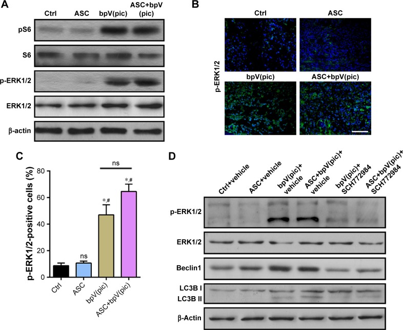
bpV(pic) promotes autophagy and attenuates apoptosis through activation of ERK1/2 signaling.
Notes: (A) Immunoblot analysis of pS6, S6, p-ERK1/2, ERK1/2, and β-actin levels in RNSCs with different treatments. n=3 independent experiments. (B) Representative immunofluorescence images of p-ERK1/2 in spinal cords from SCI rats from the different groups. Scale bar =50 µm. (C) Quantification of the p-ERK1/2-positive cells in (B). n=5. (D) Immunoblot analysis of p-ERK1/2, ERK1/2, Beclin1, LC3B, and β-actin levels with vehicle (Ctrl), ASC only, bpV(pic), ASC combined with bpV(pic), bpV(pic) plus SCH772984 or ASC combined with bpV(pic) plus SCH772984. n=3 independent experiments. *In comparison with control group, P<0.05. #In comparison with ASC group, P<0.05. ns = not significant. All data are shown as the mean ± SD.
Abbreviations: ASC, acellular spinal cord; bpV(pic), bisperoxovanadium; LC3B, light chain 3B; RNSCs, rat neuron stem cells; SCI, spinal cord injury.

