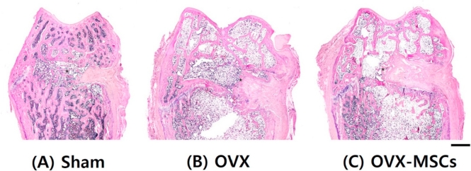
Fig. 3.
Histological observations. Hematoxylin and eosin-stained histological morphology of the entire area of distal femurs 8 weeks after surgery. (A) Sham operation. (B) Ovariectomy. (C) Mesenchymal stem cells injected after ovariectomy. Asterisk, new bone. Scale bar is 1 mm. OVX, ovariectomized; OVX-MSCs, OVX-mesenchymal stem cells.

