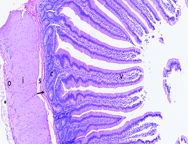
Figure 1a.

The histologic section of microscopically normal jejunum from an adult male rhesus macaque has elongated, finger-shaped villi (V) covered by an uninterrupted layer of columnar epithelial cells, and crypts (C) containing proliferative cells that replenish the superficial epithelial cells as they migrate up the villus and are eventually lost into the intestinal lumen. The muscularis mucosa (arrow) is a thin layer of smooth muscle that separates the mucosa from the underlying submucosa (s). The external muscular layer (‘muscularis externa’), divided into outer longitudinal (o) and inner circular (i) layers, serves to propel ingesta through the intestine via the process of peristalsis. The serosa (*) is thin and contains a sparse amount of fibrillar collagen. The loose connective tissue (‘lamina propria’) in the center of villi and surrounding the crypts contains a moderate number of leukocytes, including lymphocytes, macrophages, and occasionally a small number of neutrophils and eosinophils. The superficial epithelial layer of villi contains widely spaced leukocytes that are predominately T cells. Note the overall length of the villi, and the relative ratio of villus length to crypt depth. Fragments of villus epithelium in the intestinal lumen at the right side of the image are a result of tissue handling during specimen collection and gross trimming. H&E stain, 10x objective magnification.
