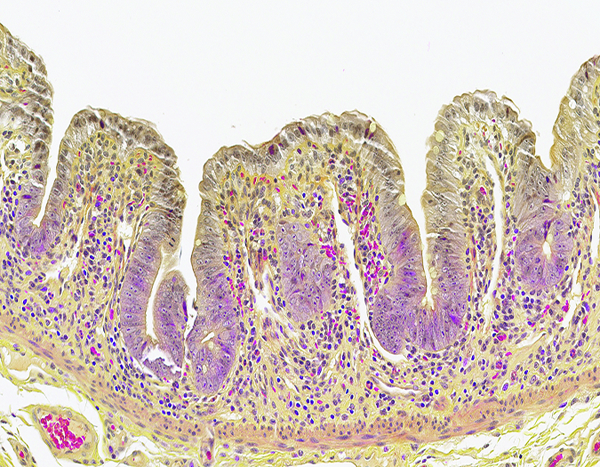
Figure 5c.

The jejunum of a male rhesus macaque collected at day 10 following irradiation at 12 Gy has severely blunted villi with proliferative crypt cells extending up the villi to the superficial mucosal surface. Note the absence of Paneth cells, as demonstrated in the following image. Erythrocytes within blood vessels and scattered in the lamina propria are stained a bright magenta color. Lendrum stain, 20x objective magnification.
