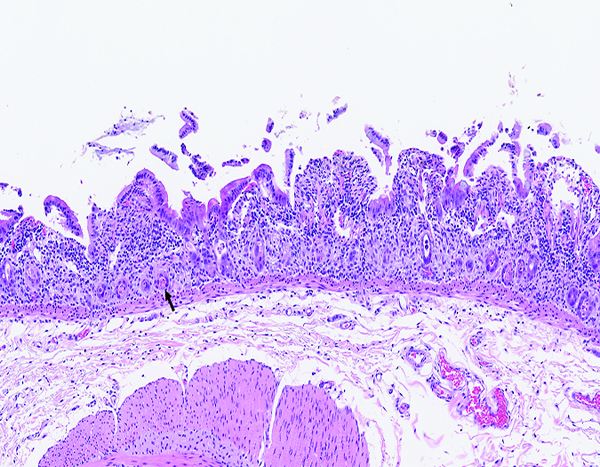
Figure 1c.

The jejunum of a male rhesus macaque collected at day 6 following irradiation at 12 Gy has markedly shortened villi. The luminal surface covered by attenuated epithelial cells, some of which are being lost as cellular rafts into the intestinal lumen. Few cellular elements remain in crypts (arrow). The leukocyte population in the lamina propria appears somewhat more prominent than in the normal jejunum, but some of this appearance may be due to contraction of the intestinal mucosa that results in visual concentration of pre-existing leukocytes. H&E stain, 10x objective magnification.
