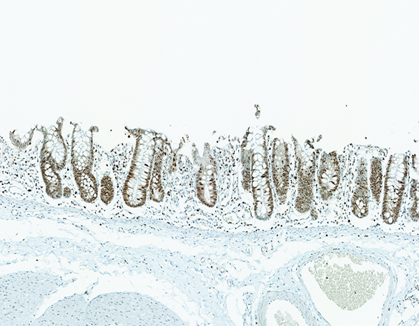
Figure 8f.

The colon of a male rhesus macaque collected at day 17 following irradiation at 11 Gy has extensive proliferative activity in crypts (*). Note the superficial zone of normal, nonproliferative epithelial cells that are visible in the colon of the naïve control animal (above) is not present in this image. Ki67 IHC stain, 10x objective magnification.
