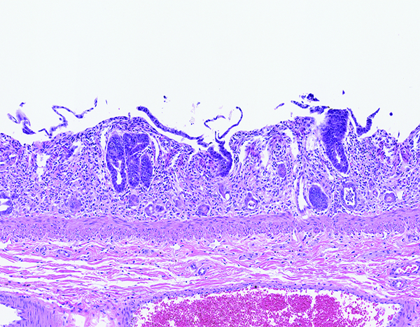
Figure 1d.

The jejunum of a male rhesus macaque collected at day 7 following irradiation at 10 Gy has no discernible villi, but multiple crypts are filled with deeply basophilic proliferative epithelial cells. Compare this image to the following image from an animal exposed to 12 Gy irradiation and necropsied on day 8. This comparison suggests crypt epithelial recovery was established sooner in animals that received the lower irradiation dose. H&E stain, 10x objective magnification.
