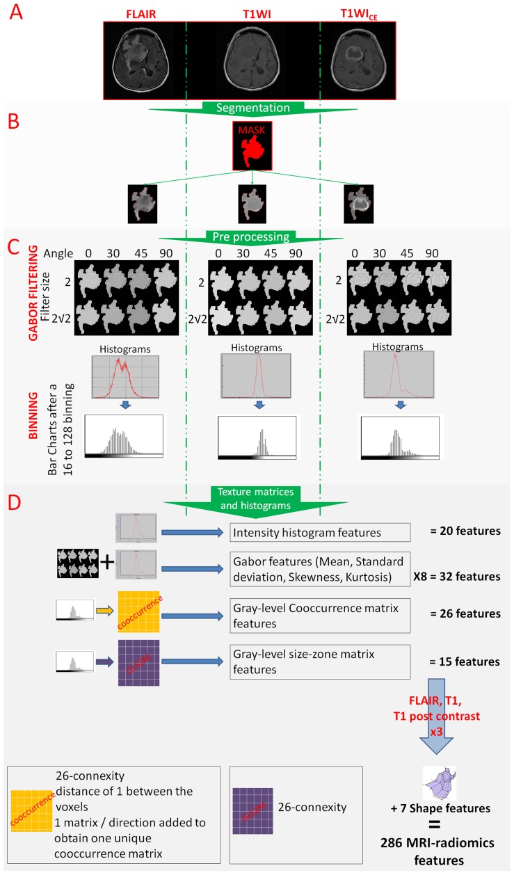
Figure 2. From the image acquisition to the radiomics features extraction.
(A). Acquisition of T1WI, T1WICE and FLAIR sequences. (B). Image segmentation using simultaneously the information from the 3 sequences (in red) and reporting this segmentation on all sequences. (C). Image post treatments: Gabor filtering and binning. (D). Radiomics feature extraction.

