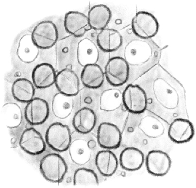
Figure 2.

(Table IV.) Cross-cut section of the content periphery of a seminiferous tubule in which [spermatids] are in their second [(elongating)]developmental stage, seen from the outside to the inside. Clear nuclei belong to [Sertoli cells]; under the mosaic lines formed by the basal surface of [Sertoli cells], it is possible to see first stage [spermatocytes]. 480/1. (This represents a stage IX–XI tubule [Perey, Clermont, and Leblond Amer J Anat (1961)], based on the presence of elongating spermatids; at these stages, the first stage spermatocytes would be leptotene.)
