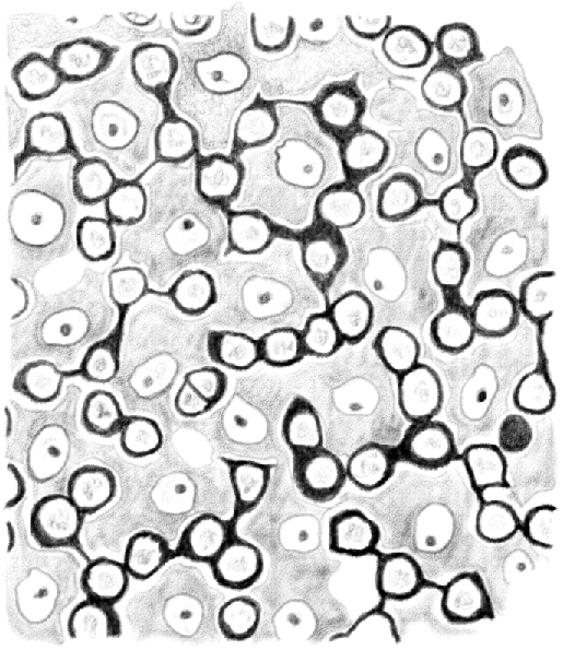
Figure 6.

(Table IV.) Cross-cut section of the content periphery of a tubule in which [spermatids] are approaching the end of their third [(condensing)] developmental stage. Second stage [intermediate (In) and type B spermatogonia] dividing and penetrating between the basal surface of [Sertoli cells] are shown. 480/1.
