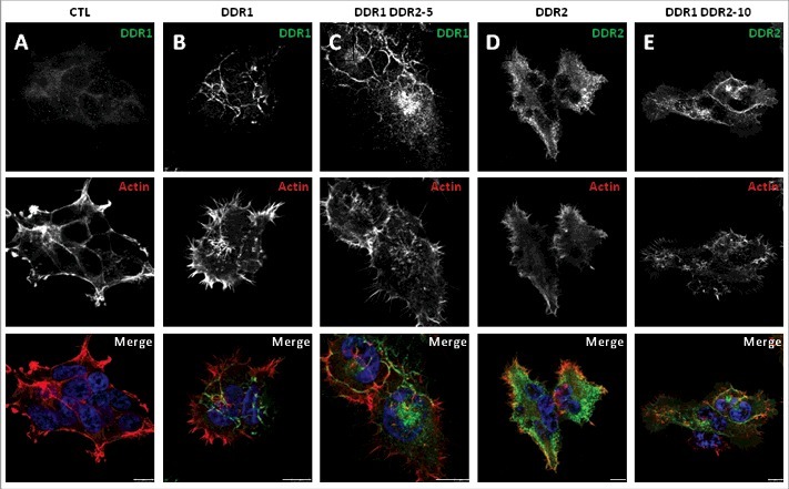
Figure 7.

Immunocytofluorescence localization of DDR1, DDR2 and filamentous actin in the different HEK 293T cell populations cultured in presence of collagen I. Cells were cultured for 24 h on glass coverslips coated with collagen I, fixed and stained with DDR1 or DDR2 antibody (CST, green) as indicated and with phalloidin (filamentous actin, red). Images were captured using a confocal microscope (Leica SP5). Bar: 10 µm. N = 3.
