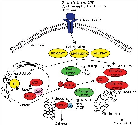
Figure 1.

Schematic diagram of MCL-1 regulation and function. MCL-1 is induced by a variety of extracellular receptor tyrosine kinases (RTKs) (purple), growth factors, stress induced cytokines and hormones resulting in the activation of intracellular signals (yellow) and inducing the transcription of MCL-1 mRNA by target transcription factors (TF). MCL-1 activity and stability is regulated by E3 ubiquitin-ligase and protein kinase induced phosphorylation (P) during post-translational processing. The best recognized function of MCL-1 is its role in maintaining cell survival via interaction with the intrinsic apoptotic machinery at the mitochondria. MCL-1 can also participate in the regulation of mitochondrial structure and function, cell cycle (CellC) and DNA damage mechanisms. In diseased and damaged cells, MCL-1 can be ubiquitinated (U) and targeted for degradation at the proteasome resulting in cell death.
