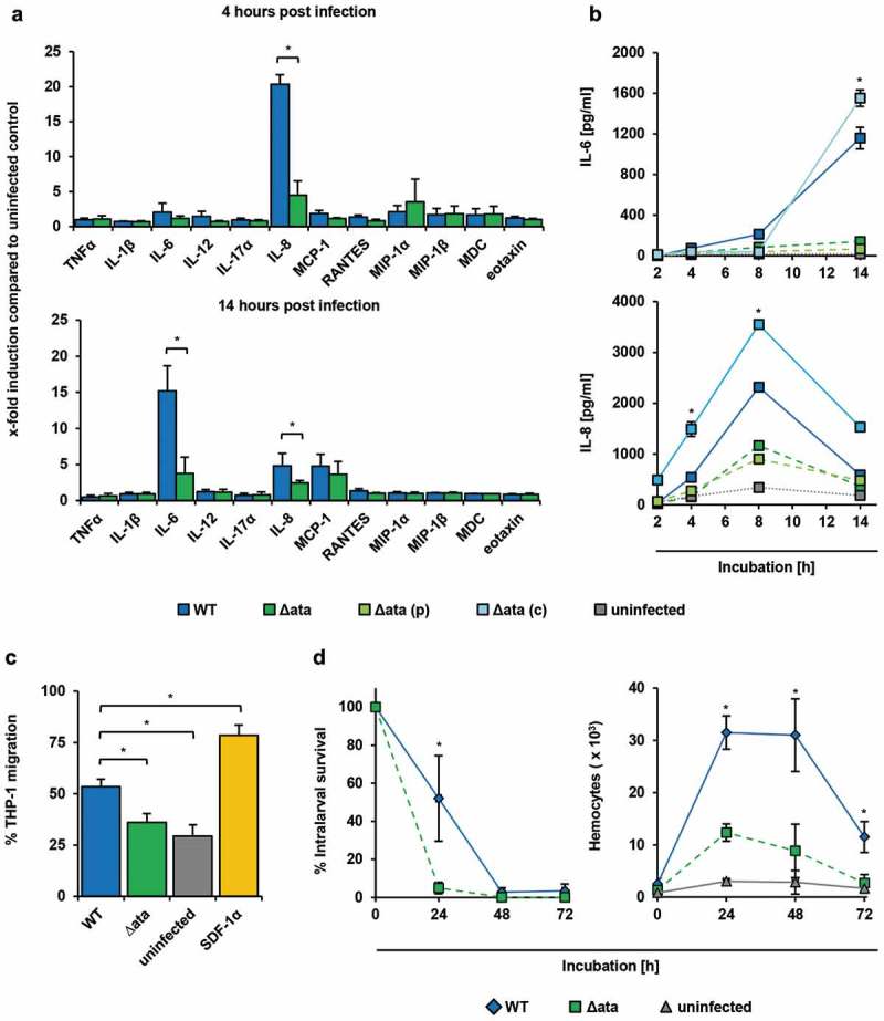
Figure 3.

A. baumannii induce inflammatory response in endothelial cells and supports migration of immune cells. (a+b) Ata-mediated induction of inflammatory cytokines upon infection of HUVECs. HUVECs were incubated with A. baumannii (MOI 1) for the indicated time points, and levels of secreted chemokines and cytokines were determined in the supernatant by ELISA. Values are means ± SEM of six independent experiments; *, p < 0.05. (c) Infection of HUVECs with A. baumannii supports transmigration of THP-1 cells. Sterile filtered supernatants of A. baumannii infected HUVECs (MOI 1, 14 h) were used as chemoattractant for analyzing transmigration of THP-1 cells. Monocytes (5 × 10⁵) were placed into the upper part of a cell culture insert (pore size: 8 µm) and allowed to migrate for 16 h towards the chemoattractant in the lower part of the well. THP-1 cells were counted using trypan blue staining and a hemocytometer. (d) Survival of A. baumannii in G. mellonella and its contribution to activate hemocytes within the larvae. Caterpillars were infected with a sub-lethal dose of A. baumannii (1 × 10⁵ bacteria). For analyzing the survival of A. baumannii, larvae were homogenized at the indicated time points and serial dilutions were plated onto Endo agar (BD) for CFU enumeration. To investigate the activation of hemocytes, larvae were homogenized and centrifuged in a cell filter containing tube to separate the hemolymph. Samples were mixed with 100 µL of trypsin-EDTA (0.05%) and stained with trypan blue, immediately. Hemocytes were enumerated using a hemocytometer. In (c)+(d), values are means ± SD of three independent experiments; *, p < 0.05.
