Fig. 1.
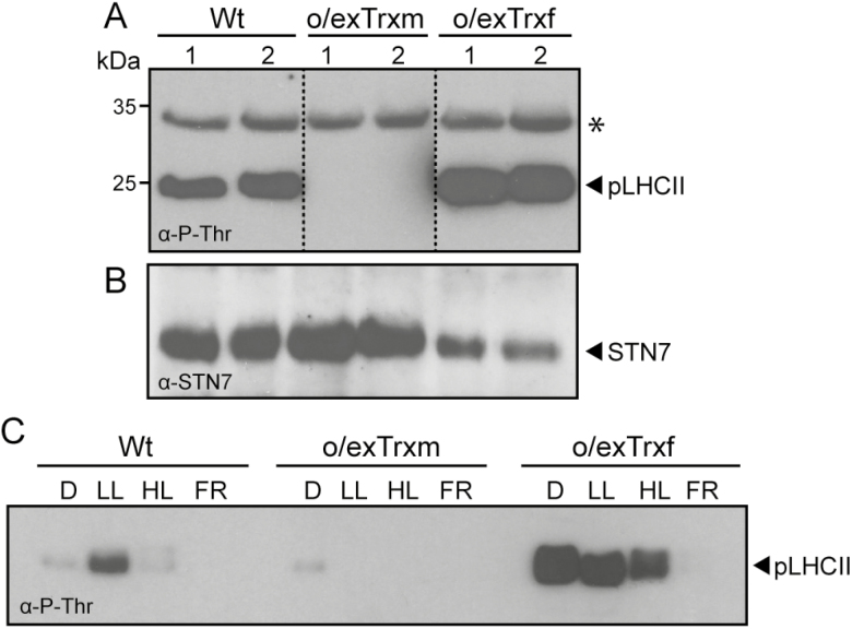
Effect of Trx m or f overexpression in tobacco chloroplasts on thylakoid protein phosphorylation and STN7 accumulation. (A) Thylakoid protein phosphorylation in Wt, o/exTrxm, and o/exTrxf plants sampled during the light (16 h at 80 μmol m−2 s−1) period. Thylakoid proteins (15 µg) were separated by SDS–PAGE (15%+6 M urea), transferred to a PVDF membrane, and immunoblotted with a phosphothreonine antibody. Phosphorylated LHCII (pLHCII) proteins are indicated. The asterisk represents phosphorylated PSII core proteins (D1 or D2). Molecular weights (kDa) are indicated on the left. (B) STN7 protein accumulation in the samples described in (A). The PVDF membrane was probed with an antibody raised against STN7. (C) LHCII phosphorylation pattern under different light regimes. Wt, o/exTrxm, and o/exTrxf plants were placed in darkness (D) for 8 h and then transferred for 2 h to low light (LL; 80 μmol m−2 s−1), followed by exposure to high light (HL; 800 μmol m−2 s−1) or far red light (FR) for 1 h. At the end of light treatments, the LHCII phosphorylation of isolated thylakoids was analyzed by western blot.

