Figure 1.

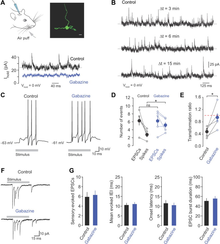
Inhibition regulates the sensory-evoked input–output function of granule cells. A, Whole-cell recordings were made from granule cells in vivo during brief air-puff stimulation of the ipsilateral upper lip area and whiskers (gray arrow). Morphological identification of individual granule cells was achieved by biocytin labeling through the recording electrode and subsequent staining with streptavidin Alexa Fluor 488. Scale bar, 5 μm. Bottom, Voltage-clamp recordings at 0 mV in the absence (control) and presence of gabazine (500 μm). B, Voltage-clamp recordings of IPSCs recorded at 0 mV at 3, 6, and 15 min after “break-in.” C, Representative current-clamp recordings of action potentials evoked in response to whisker stimulation in control and gabazine (500 μm). D, The relationship between evoked mossy fiber input (EPSCs) and granule cell output (spikes) in control and in the presence of gabazine (n = 5). E, Transformation ratio (number of evoked spikes/number of evoked EPSCs) in control and gabazine. F, Sensory-evoked EPSCs recorded from a granule cell held at −70 mV in control and in the presence of gabazine. G, Average number of EPSCs, evoked IEI, onset latency, and sensory-evoked burst duration in control and gabazine (n = 12).
