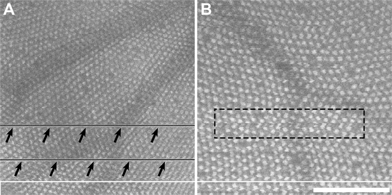
Fig. XX.1.

Disruptions in en face images of the cone mosaic derived from OCT volume scans. (A) Significant gill movements during the process of scanning result in missing data in the en face image (arrows). (B) Subtler gill movements and/or errors in contour placement can result in localized distortions (dashed rectangle). While these distortions may not affect the ability to identify the cones in the images, they would affect measurements of mosaic geometry (Cooper et al. 2016b). In addition, shadowing from overlying retinal vasculature was seen in many images. Scale bar = 100 μm.
