
Figure 6. Orientation-selected Q-band Mims-ENDOR spectra of the paramagnetic intermediate with magnetic nuclei (2H7 and 15N1) from the substrate DTB.
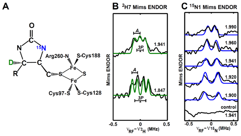
A, Depiction of the intermediate with the incorporated nuclei 2H7 marked in green, and 15N1 marked in blue. The geometry of the intermediate is adapted from the DFT model given in Figure 7A & Figure 8B. B, Mims-ENDOR spectra of 2H7-DTB-labled intermediate. C, Mims-ENDOR spectra of 15N1-DTB-labled intermediate.
Experimental parameters: 10 K, microwave pulse π/2 = 12 ns, τ = 240 ns for 2H7 and 300 ns for 15N1, RF pulse = 20 μs. Simulation parameters: g = [1.993, 1.941, 1.847]; A(15N1) = [−0.14, −0.25, −0.39] MHz, Euler angle = [0, 10, 0]°; A(2H7) = [0.03, 0.03, 0.09] MHz, P(2H7) = [−0.02, −0.08, 0.10] MHz, Euler angle = [0, 150, 0]°. All the experimental spectra are in black, while the simulated spectra are colored. The BioB enzyme used to generate the intermediate sample is natural abundant and (guanidino-15N2)-arginine enriched for 15N1-DTB-labeled and 2H7-DTB-labeled intermediate, respectively.