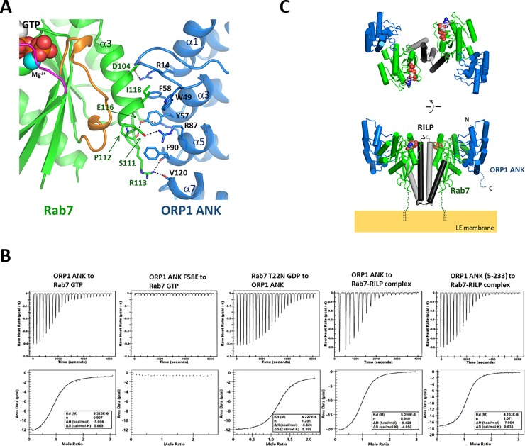
Fig 3. Formation of the ORP1-Rab7-RILP complex.
(A) Binding interface of the ORP1 ANK and Rab7. The residues forming the interface are shown in stick models. The hydrogen bonds are shown in dashed lines. (B) Measurement of binding affinity of ORP1 ANK to Rab7 by isothermal titration calorimetry. The purified Rab7 or Rab7-RILP complex with the concentration of 0.1 mM was titrated with 1 mM of ORP1 ANK. Rab7 was either loaded with GTP or GDP. (C) Overall structure of the ORP1 ANK-Rab7-RILP complex. The structure was constructed by combining the structures of Rab7-RILP (PDB id: 1YHN) and ORP1 -Rab7 (this study) using PyMOL (https://pymol.org). The missing C-terminal residues (186–203) of Rab7 were indicated with dotted lines. The C-terminal prenyl groups which anchor Rab7 onto the endosomal membrane are shown in black lines.

