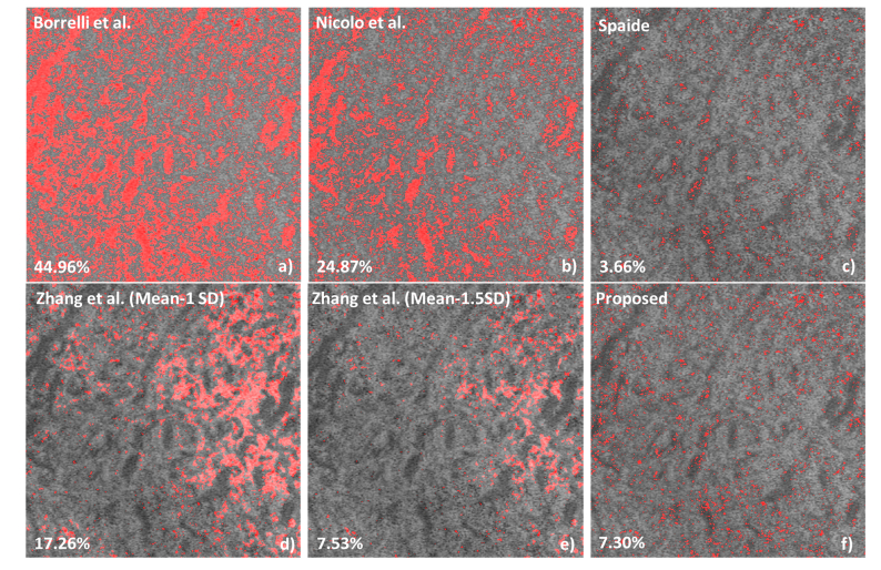
Fig. 6.
The demonstration of applying different flow void segmentation algorithms on a 3 × 3 mm2 choriocapillaris image centered at the fovea. (a) Borrelli et al. [34] used the global threshold method where the threshold level was set to be the mean intensity of the avascular outer retinal layer. (b) Nicolo et al. [24] applied Otsu’s thresholding method. (c) Spaide [26]. used Phansalkar local threshold method. (d-e) Zhang et al. [29] used the structural image to compensate the angiogram, and then used mean-1SD and mean-1.5SD to binaries the compensated images.

